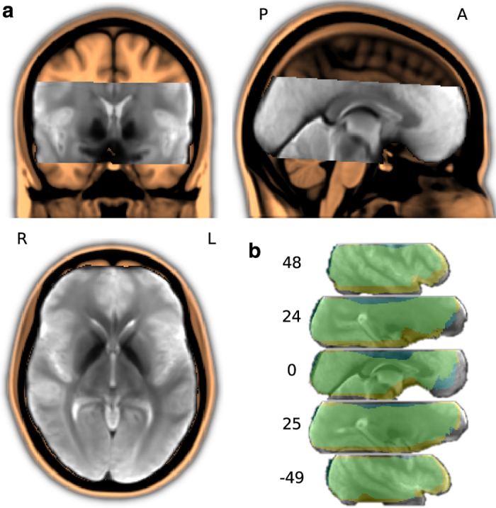
Figure 2.

Field-of-view for the fMRI data acquisition. (a) Custom T2* EPI group template (in gray), linearly aligned to and overlaid on the MNI152 T1-weighted head template (in amber). The EPI template was created by an iterative procedure with four linear and ten non-linear alignment steps out of one sample volume per run and brain (a total of 152 images; images from participant 10 were excluded; see Table 3, slice cut point: anterior commissure at MNI 0,0,0 mm). (b) Intersection masks after linear and non-linear anatomical alignment of all mean volumes for all individual fMRI runs across all participants. The linear intersection mask is depicted in blue, non-linear in yellow (overlap in green). Coordinates are in MNI millimeters.
