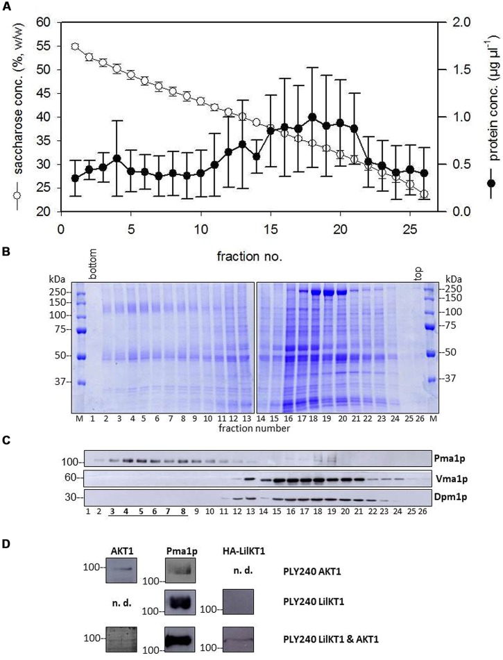
FIGURE 4.
Co-sedimentation of LilKT1 with yeast organelle markers. (A) Distribution of membrane protein and sucrose concentration in a continuous sucrose gradient collected after 16 h centrifugation (n = 5 ± S.D.) (B) Typical pattern of membrane proteins along the sucrose gradient. 30 μl of sucrose-adjusted fraction loaded per lane. (C) Typical distribution of plasma membrane (Pma1p), ER (Dpm1p), and vacuole (Vma1p) marker enzymes in the sucrose gradient. (D) Plasma membrane fractions (3–8) of three gradients were pooled, proteins were precipitated and transferred to NC membranes after SDS-PAGE. The proteins AKT1, Pma1p and the HA-tag of LilKT1 were detected with respective antibodies in the pooled fractions of PM fractions (underlined) of PLY240 yeast mutants expressing AKT1 or LilKT1 alone and the combination of AKT1 and LilKT1 (n.d., not determined).

