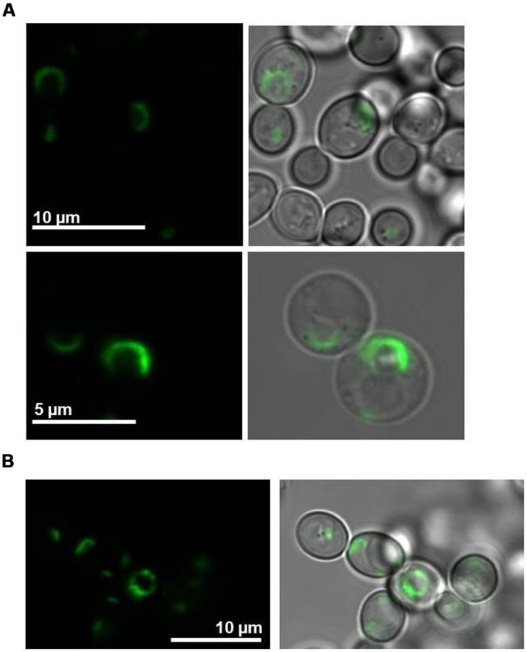
FIGURE 5.
Localization of fluorescence-tagged LilKT1 in yeast protoplasts. (A) N-terminally GFP fused LilKT1 was expressed in yeast cells and localizes in structures surrounding the nucleus (ER). Upper panel: yeast cells, lower panel: yeast cell protoplasts. Fluorescence images are on the left and bright field images merged with fluorescence images on the right. (B) Localization of GFP::LilKT1 co-expressed with AKT1 in yeast cells. Co-expression with AKT1 shows the localization in similar compartments as expression of LilKT1 alone (A).

