
Figure 3.
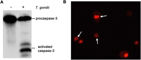
Apoptosis detected by Western blotting and nucleus staining. (A) The C17.2 cells were collected after they were co-cultured with 5 × 10⁵ RH tachyzoites for 24 h, and the activity of caspase-3 was detected by Western blotting; (B) The nuclei of the apoptotic cells were stained by propidium iodide. The arrows represent a shrinking nucleus or apoptotic body.