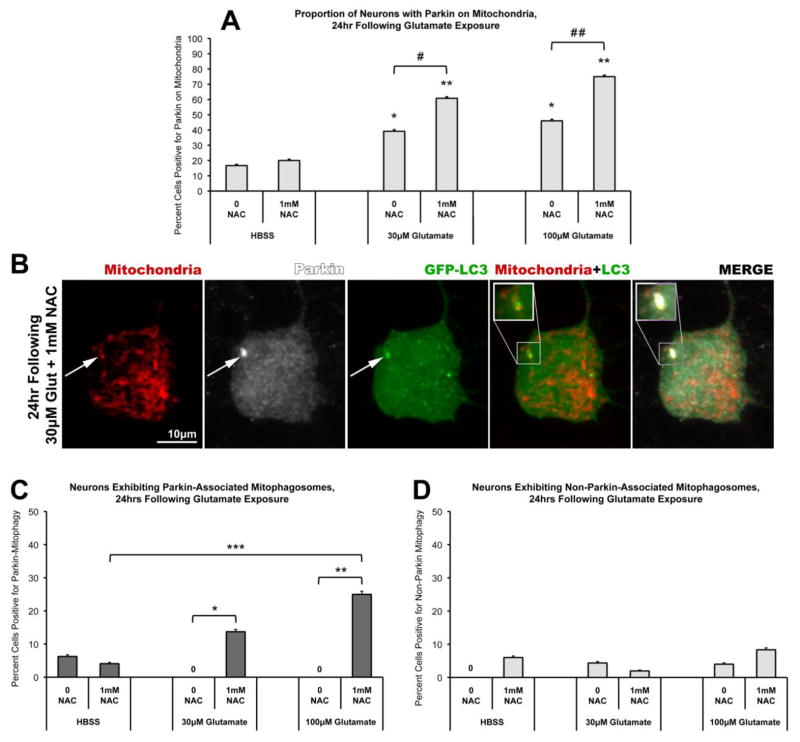
Figure 6. NAC co-treatment facilitates Parkin-associated mitophagy following glutamate exposure.
Rat cortical neurons were co-transfected with full-length human Parkin, mtDsRed2, and GFP-tagged LC3 (GFP-LC3) at DIV6. At DIV15, neurons were exposed to a brief (10min) treatment with either HBSS media or Glutamate (30μM or 100μM), with or without 1mM NAC, followed by a 24hr incubation period in culture media. Cells were then assessed by confocal analysis for localization of Parkin immunofluorescence relative to mtDsRed2 fluorescence (Mitochondria) and GFP immunofluorescence (GFP-LC3). (A) Glutamate-induced Parkin localization to mitochondria was observed 24hr following both 30μM and 100μM glutamate exposure (46–51 individual cells per condition, across 4 independent neuronal preparations; * = p<0.05 from HBSS, 0 NAC; ** = p<0.05 from HBSS, 1mM NAC; +/− SEM). Parkin localization was significantly increased by 1mM NAC co-treatment with glutamate, as compared to glutamate alone (# = p<0.05 between 30μM glutamate, 0 NAC and 30μM glutamate, 1mM NAC; # # = p<0.05 between 100μM glutamate, 0 NAC and 100μM glutamate, 1mM NAC; +/− SEM). (B) Instances of Parkin-associated mitophagosome formation were observed as co-localization of both Parkin (white) and GFP-LC3 (green) accumulations on a mitochondrion (red) (inset). (C) Graph of the observed percentage of neurons exhibiting Parkin-associated mitophagosomes 24hr following glutamate exposure with or without NAC co-treatment (46–51 individual cells per condition, across 4 independent neuronal preparations; * = p<0.05 between 30μM glutamate, 0 NAC and 30μM glutamate, 1mM NAC; ** = p<0.05 between 100μM glutamate with, 0 NAC and 100μM glutamate, 1mM NAC; *** = p<0.05 between HBSS, 1mM NAC and 100μM glutamate, 1mM NAC; +/− SEM). (D) Instances of mitophagosome formation not associated with Parkin were also observed in these experiments (measured as colocalization of GFP-LC3-labeled autophagosomes with mitochondria, but not containing Parkin). The proportions of cells exhibiting this mitophagy were not significantly different from controls nor significantly influenced by NAC co-treatment (+/− SEM).

