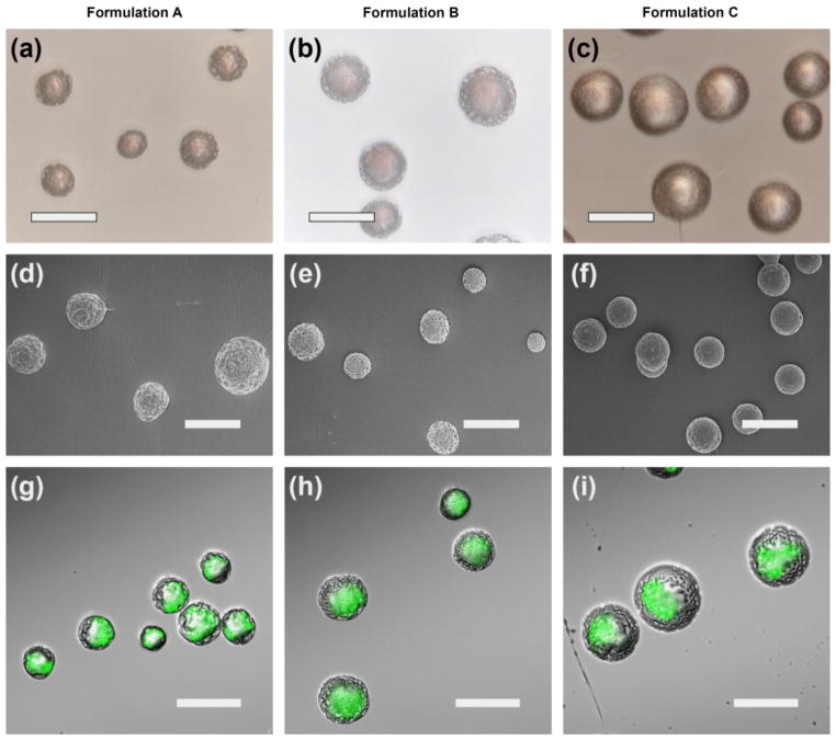
Fig. 11.
Transmitted light microscopy (a–c), scanning electron microscopy (SEM) (d–f), and laser confocal scanning microscopy (g–f) of core-shell microparticles produced using coaxial electrohydrodynamic atomization The green light represent the location of core structure (PLGA) surrounded by shell (PDLLA) (reprinted from Xu et al., 2013a, with permission from Elsevier).

