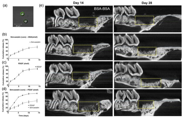
Fig. 8.
(a) Fluorescent image illustrating core-shell structure of microspheres. (b–d) The in vitro release profiles of different formulations. (e) Micro-CT characterization of a sagittal slice crossing the mid-points of the maxillary second molar and third molar. BSA-BSA: BSA(core)-BSA(shell). (reprinted from Chang et al., 2013, with permission from Elsevier)

