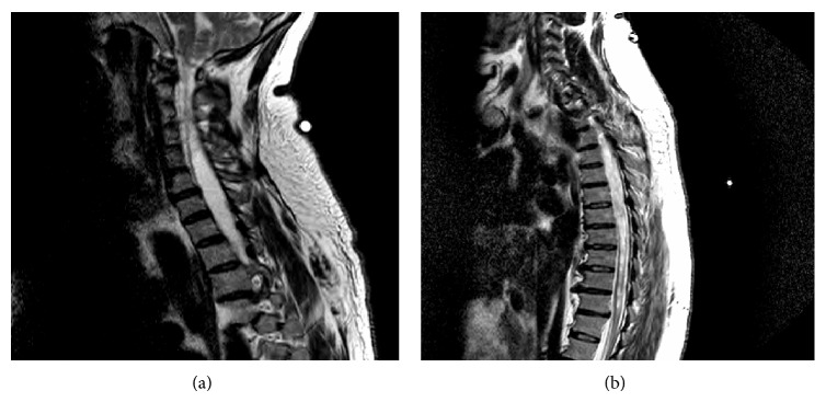
Figure 3.

(a) MRI of the cervical spine showing a 9 mm inferior herniation of the cerebellar tonsils through the foramen magnum (Type I Chiari Malformation), with extensive cervical and superior thoracic spinal cord syringohydromyelia. (b) Thoracic spine MRI revealing extensive syringohydromyelia involving the entire thoracic spinal cord. The syringohydromyelia is more prominent in the superior midportion of thoracic spinal cord. T11-T12 central disk protrusion with thecal sac compression and slight ventral spinal cord compression.
