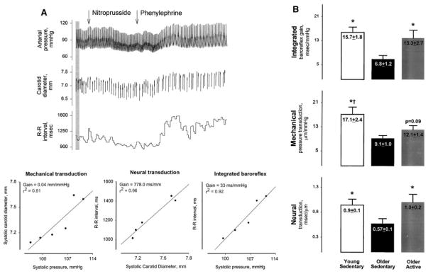
Fig. 7.
Analysis of vascular and neural components of cardiovagal baroreflex sensitivity in humans. Panel A shown are measurements of arterial pressure, carotid artery diameter, and R-R interval from one subject during nitroprusside- and phenylephrine-induced decreases and increases in arterial pressure, respectively. The calculated slopes of relationships between systolic carotid diameter and systolic pressure (left), R-R interval and systolic diameter (middle), and R-R interval and systolic pressure (right) denote the sensitivity of the mechanical component, neural component, and integrated baroreflex, respectively. Panel B shown are data from young sedentary, older sedentary, and older active subjects. Both mechanical and neural components of the reflex were significantly decreased in the older sedentary group. Baroreflex sensitivity was preserved in active older individuals, an effect attributed primarily to a positive effect of activity on the neural component of the reflex. Reproduced with permission from Hunt et al. 2001 [112, 113]

