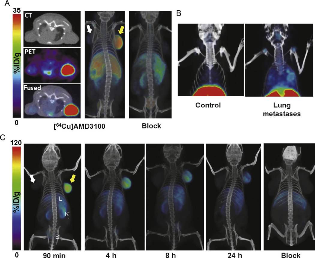
Figure 2.3.
Volume rendered whole body PET/CT images of CXCR4 expression in subcutaneous NOD–SCID mice bearing U87-stb-CXCR4 (yellow arrow) and U87 control (white arrow) tumors (A) and MDA-MB-231-derived lung metastases (B) following injection of 300 µCi of 64Cu-AMD3100; U87-stb-CXCR4 (yellow arrow) and U87 control (white arrow) tumors following administration of 250 µCi of 64Cu-AMD3465 or a 25-mg/kg AMD3465 blocking dose followed by 250 µCi of 64Cu-AMD3465 (C). Images (A,B) were recreated from Nimmagadda et al. (2010) and (C) from De Silva et al. (2011).

