
Figure 6. PD-L1 expression in tumor tissue samples and its correlation with recurrence free survival in nasopharyngeal carcinoma patients.
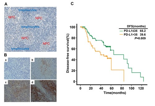
(A) Histological features of NPC: Nasopharyngeal carcinoma cells (red) were surrounded by infiltrating lymphocytes (blue). (B) PD-L1 expression in nasopharyngeal carcinoma sample (a. negative staining b. weak staining c. moderate staining d. strong staining). (C) Disease-free survival in nasopharyngeal carcinoma patients stratified by the expression level of PD-L1.