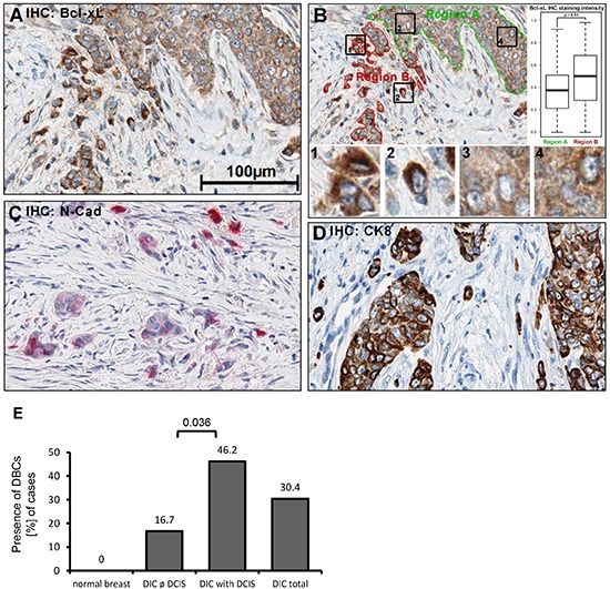
Figure 3. Bcl-xL protein levels are increased in human breast cancer cells that are dispersed in the desmoplastic stroma.

Immunohistochemistry (IHC) of human tissue, fixed in PBS-buffered formalin (4%), was embedded in paraffin. 1.5μm sections were treated with boric-acid/EDTA buffer for antigen-retrieval, followed by incubation with primary antibodies, secondary peroxidase-coupled antibodies, and diaminobenzidine (DAB). A more extensive array of staining is shown in Supplemental Fig. S3. (A) Detection of Bcl-xL in breast cancer samples. (B) Quantification of DAB precipitate color-intensity in the two highlighted regions (borders green, red); boxplots of respective intensities. 1′-′4′: Magnified details as indicated. Statistical testing was performed using Student´s T-test. (C) N-cadherin IHC (Fast Red). (D) Cytokeratin 8 IHC. B-D: 400x magnification. (E) Presence of dispersed, Bcl-xL enhanced cells (DBCs) in correlation to the presence and absence of ductal carcinoma in situ (DCIS), n = 56.
