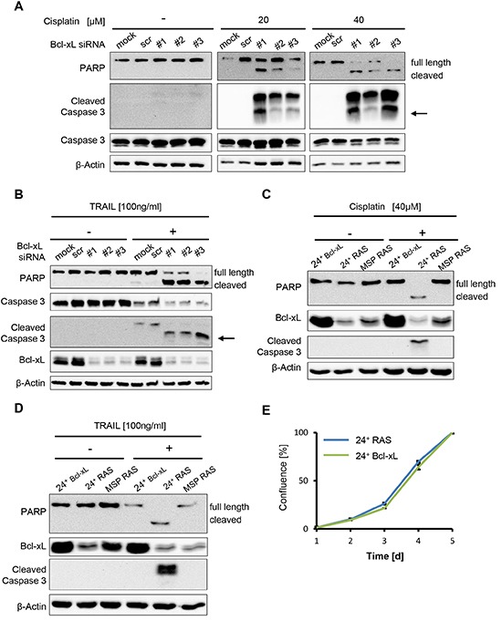
Figure 4. Bcl-xL levels determine apoptosis but not cell proliferation in HMLE RAS cells.

(A, B) MSP RAS cells were depleted of Bcl-xL by siRNA and treated with 20μM and 40μM cisplatin for 16h (A), or with 100ng/ml Trail in the presence of 20μg/ml of the inhibitor of protein synthesis cycloheximide (CHX) for 6h (B). Cell lysates were analysed by immunoblotting. Untreated (mock) and scrambled (scr) siRNA-transfected cells were used as controls (see also Supplemental Fig. S4B). An arrow is indicating the molecular weight of cleaved caspase 3 (17kDa). A longer exposure for non-treated cells was used to ensure that cleaved Caspase 3 would have been detected if present. (C, D) 24+ Bcl-xL and HMLE RAS cells were treated with 40μM cisplatin for 16h (C) or 100ng/ml Trail in the presence of 20μg/ml CHX for 6h (D), followed by immunoblot analysis. (E) Proliferation of 24+ RAS and 24+ Bcl-xL overexpressing cells was monitored for 5d. Cell confluence was determined daily using automated light microscopy with quantitative image analysis (Celigo cytometer).
