
Figure 1. Acidosis increases invasive capacity and NF-κB activity.
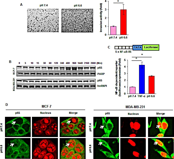
(A, left) MDA-MB-231 cells were cultured at pH 7.4 or pH 6.6 for 48 hours and then assessed in vitro in regular medium using Matrigel invasion chambers. (A, right) Fold induction in the number of invaded cells at pH 6.6 as compared to those cultured at pH 7.4. (B) Time-dependent effects of acidic environment on NF-κB p65 nuclear translocation. Cells were cultured at pH 6.6 for the indicated time, nuclear extracts were prepared and assayed for p65 content by Western blotting. (C) Acidic environment induces NF-κB-dependent reporter gene expression in MCF-7 cells. Cells were transiently transfected with a plasmid in which a luciferase reporter carries 5 copies of NF-κB response elements (5 × NF-κB-RE) in front of minimal promoter (TATA). Cells were then cultured at pH 7.4 or pH 6.6 for 12 hours and luciferase activity was measured using whole cell lysate. Where indicated, the cells cultured in normal medium were exposed to 0.5 nM TNF-α for 12 hours. (D) Cellular localization of NF-κB p65 in breast cancer cells under acidic environment. Cells were cultured at pH 7.4 or pH 6.6 for 1 hour and analyzed for p65 localization by immunefluorescent staining. *, p< 0.05.