
Figure 4. The acidosis-induced activation of NF-κB and AKT is mediated through ROS generation.
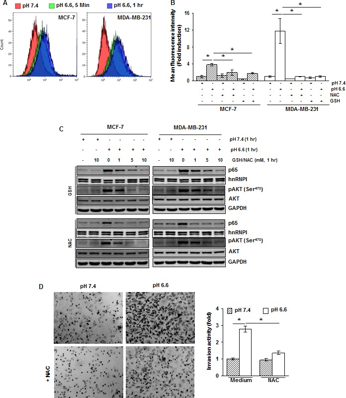
(A) Cells were cultured at pH 7.4 and pH 6.6 for 5 minutes and 1 hour. The intracellular ROS levels were measured using DCFH-DA by flow cytometry. (B) ROS generated by acidosis is suppressed by NAC and GSH. Cells were treated with NAC (10 mM) or GSH (10 mM) for 1 h, washed off, and then cultured at pH 7.4 or pH 6.6 for 1 hour. The intracellular ROS levels were measured using DCFH-DA by flow cytometry. (C) Cells were treated with indicated concentrations of GSH or NAC for 1 hour, washed off, and then cultured at pH 7.4 and pH 6.6 for 1 hour. The nuclear extracts and whole-cell extracts were analyzed by Western blotting for p65 and pAKT, respectively. (D) NAC suppresses acidosis induced invasion activity of MDA-MB-231 cells. Cells were pretreated with NAC (10 mM) for 1 hour, washed off, and then cultured at pH 7.4 or pH 6.6 for 48 hours. Invasion activity was measured using Matrigel invasion chambers.*, p< 0.05.