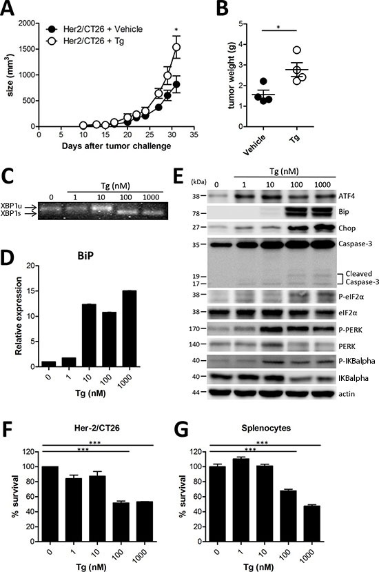
Figure 1. ER stress induced by Tg accelerated tumor growth.

(A) BALB/c mice were injected s.c. with 106 HER2/CT26 cells per mouse, and 100 μg/kg of Tg was administered i.p. every day before tumor challenge. Tumor growth was monitored (n = 5). (B) tumor weight at 4 weeks after HER2/CT26 injection (n = 4). Graphs show mean ± SEM. *p < 0.05, ***p < 0.001 compared with matched control group using the Student's t-test. (C) XBP1 mRNA splicing in Tg-treated HER2/CT26 cells. (D) mRNA levels of BiP from Tg-treated HER2/CT26 cells as measured by RT-qPCR. (E) immunoblot of Tg-treated HER2/CT26 cells for the PERK-eIF2α branch. (F) HER2/CT26 cells were cultured with Tg for 24 h and cell viability was analyzed. (G) splenocytes were cultured with Tg for 24 h and cell viability was analyzed. ***p < 0.001 using one-way ANOVA with Tukey's post hoc test.
