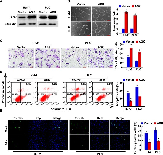
Figure 2. Overexpression of AGK promotes angiogenesis and inhibits apoptosis in HCC cells in vitro.

(A) Overexpression of AGK in Huh7 and PLC cell lines was confirmed by western blotting; α-tubulin was used as a loading control. (B) Representative images (left) and quantification (right) of tubule formation by HUVECs cultured on Matrigel-coated plates with conditioned medium collected from the indicated HCC cells. Scale bars: 200 μm. Each bar represents the mean ± SD of three independent experiments; *P < 0.05. (C) Cell migration was assessed by culturing HUVECs with conditioned media collected from the indicated HCC cells. Scale bars: 50 μm. Each bar represents the mean ± SD of three independent experiments; *P < 0.05. (D) Annexin V-FITC/PI staining of the indicated cells after treatment with cisplatin (20 μM) for 24 hours. Each bar represents the mean ± SD of three independent experiments; *P < 0.05. (E) Representative images (left) and quantification of TUNEL-positive cells in the indicated cells after treatment with cisplatin (20 μM) for 24 hours. Scale bars: 50 μm. Each bar represents the mean ± SD of three independent experiments; *P < 0.05.
