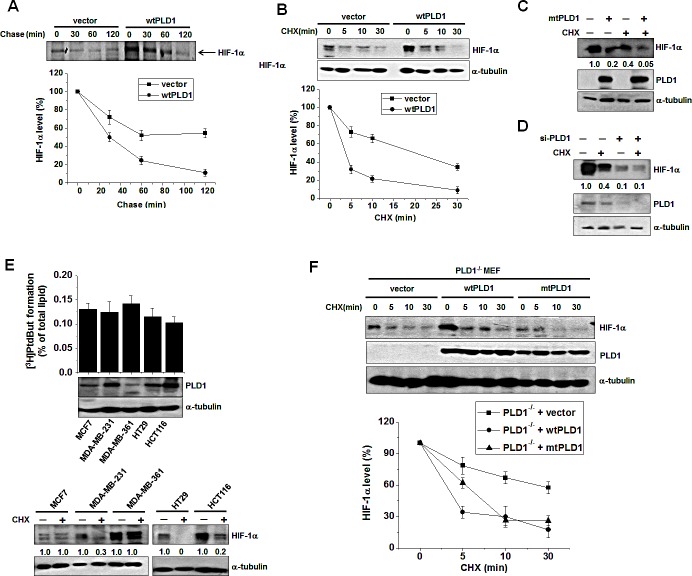
Figure 1. PLD1 plays a dual role in regulation of the cellular level of HIF-1α protein.
(A) Pulse-chase assay of HIF-1α in HEK293 cells transfected with vector or wtPLD1. The cells were pulse labeled with [35S]methionine-cysteinefor 4 h in the presence of MG132 and then chased in unlabeled medium for the indicated time. Lysates were immunoprecipitated with antibody to HIF-1α and assessed by autoradiography, after which the band intensity was quantified relative to the level of HIF-1α of no chase. Data are representative of three independent experiments. (B) Effects of PLD1 on the stability of HIF-1α. HEK293 cells were transfected with vector or PLD1 and then incubated under hypoxia (1% O2) for 4 h. The cells were subsequently reoxygenated (21% O2) and treated in parallel with CHX (100 μg/ml) for the indicated time. Lysates were analyzed by immunoblotting, after which the band intensity was quantified. The levels of HIF-1α to α-tubulin were normalized. Data are representative of three independent experiments. (C) HEK293 cells were transfected with mtPLD1 and incubated under hypoxia for 4 h, then reoxygenated by treatment with CHX for 30 min. Lysates were subsequentlyimmunoblotted using the indicated antibodies, after which the band intensity was quantified and the levels of HIF-1α to α-tubulin were normalized. Data are representative of three independent experiments. (D) HEK293 cells were transfected with PLD1 siRNA and incubated under hypoxia. The cells were then reoxygenated and in parallel and treated with CHX for 30 min, after which lysates were immunoblotted using the indicated antibodies. The band intensity was quantified and the levels of HIF-1α to α-tubulin were normalized. Data are representative of threeindependent experiments. (E) PLD activity assay and immunoblot analysis. Various cancer cells were incubated under hypoxia and then reoxygenated in parallel with treatment of CHX for 10 min. Lysates were subsequently analyzed by immunoblotting and the band intensity was quantified, after which the levels of HIF-1α to α-tubulin were normalized. Data are representative of three independent experiments. (F) PLD1-null MEF were transfected with the indicated constructs and then treated with CHX (100 μg/ml) for the indicated time. Lysates were immunoblotted with the indicated antibody. The band intensity of HIF-1α was quantified and the levels of HIF-1α to α-tubulin were normalized. Data are representative of three independent experiments.

