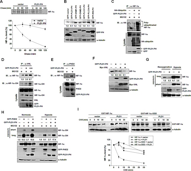
Figure 6. Disruption of the interaction of PLD1 with HIF-1α contributes to hypoxic stabilization of HIF-1α.
(A) Pulse-chase assay of HIF-1α in HEK293 cells transfected with PLD1-PH. Lysates were immunoprecipitated with antibody to HIF-1α and assessed by autoradiography, followed by quantification of the band intensity. Data are representative of three independent experiments. (B) Effect of various PH domains on the expression of HIF-1α. HEK293 cells were transfected with GFP-PH domain of various proteins and then reoxygenated. Lysates were analyzed by immunoblotting, after which the band intensity was quantified and the levels of HIF-1α to GFP-PH were normalized. Data are representative of three independent experiments. (C) Effect of PLD1-PH on the ubiquitination of HIF-1α in the presence ofMG132. The band intensity was quantified. Data are representative of three independent experiments. (D) Effect of PLD1-PH on the interaction of VHL with HIF-1α in the presence of MG132. The band intensity was quantified. Data are representative of three independent experiments. (E) Effect of PLD1-PH on the interaction of PHD2 with HIF-1α in the presence of MG132. Data are representative of three independent experiments. (F) HEK293 cells were cotransfected with PLD1-PH and/or VHLand then reoxygenated. The level of HIF-1α was analyzed by immune blot. The levels of HIF-1α to α-tubulin were quantified and normalized. Data are representative of three independent experiments. (G) IB analysis of endogenous HIF-1α by GFP-PLD1-PH under reoxygenation and hypoxia conditions. The levels of HIF-1α to α-tubulin were quantified and normalized. Data are representative of three independent experiments. (H) IB analysis of hydroxylated HIF-1α of lysates from HEK293 cells cotransfected with PHD2 and/or GFP-PLD1-PH under normoxia and hypoxia conditions in the presence of MG132. The levels of hydroxylated HIF-1α to HIF-1α were quantified and normalized. Data are representative of three independent experiments. (I) Effect of PLD1 on the stability of HIF-1α-ODD. HEK293 cells were cotransfected with the indicated constructs. CHX was added for the indicated time and lysates were analyzed by immunoblotting, after which the band intensity was quantified. The levels of HIF-1α to α-tubulin were quantified and normalized. Data are representative of three independent experiments.

