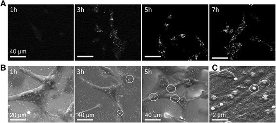
Figure 2.

Nanoparticle - cell interaction. A) Time lapse multiphoton microscopy of granulosa cells with 150 nm particles after 1 h, 3 h, 5 h and 7 h of co-incubation. B) ESEM and C) SEM images of ZMTH3 cells after different incubation times with 200 nm gold particles. B) ESEM images: After 1 h a loose dispersion of particles is visible. After 3 h the AuNP started to aggregate (ellipse). The formation of particle clusters at the membrane can be observed after 5 h. C) SEM image in a higher magnification: After an incubation time of 3 h, particles are either on the cell membrane (solid ellipse) or covered by the membrane (dashed ellipse) [24].
