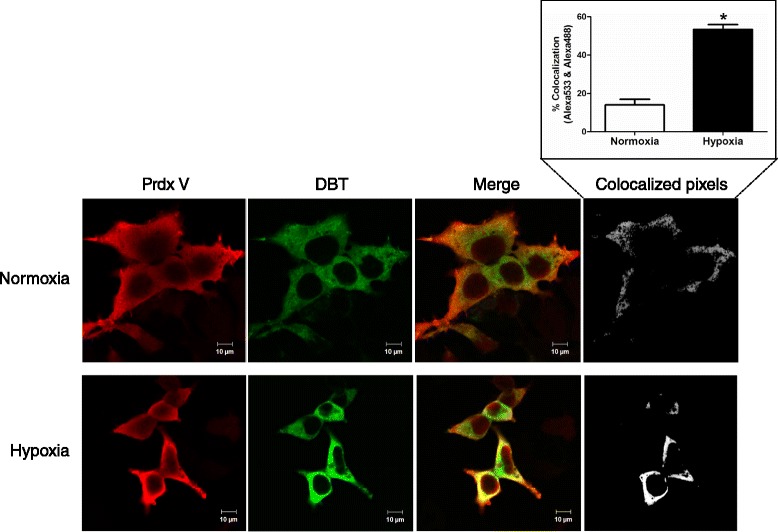
Figure 3.

Co-localization of Prdx V and DBT in normoxic and hypoxic HEK293 cells. Confocal images of costaining with antibodies to Prdx V and DBT. HEK293 cells were co-transfected with Prdx V- and DBT-expressing vectors, and then the transfected cells were exposed to hypoxic stress (1 ± 0.2%) for 6 h. Cells were fixed on polylysine slides and stained with Prdx V (red) or DBT (green) antibodies.
