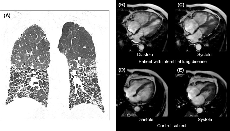
Figure 2.

Comparison of cine CMR between a patient with ILD and a control subject. (A): Chest computed tomography demonstrated diffuse fibrosis mainly located in the lower lungs in a patient with interstitial lung disease (ILD). Cine CMR showed an enlarged right ventricle (RV) and decreased RV function in an ILD patient (B: end-diastolic and C: end systolic image). In the control subject, the RV size was substantially smaller than in the ILD patient (D: end-diastolic and E: end systolic image). ILD: interstitial lung disease; CMR: cardiovascular magnetic resonance; RV: right ventricle.
