
Figs. 2A–C.
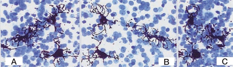
A series of rotating images of three HSCs in a liver lobule of the pig. A: 0° ; B: 120°; C: 180° Golgi silver method. The sections are stained with toluidine blue. × 350.
Official websites use .gov
A
.gov website belongs to an official
government organization in the United States.
Secure .gov websites use HTTPS
A lock (
) or https:// means you've safely
connected to the .gov website. Share sensitive
information only on official, secure websites.
A series of rotating images of three HSCs in a liver lobule of the pig. A: 0° ; B: 120°; C: 180° Golgi silver method. The sections are stained with toluidine blue. × 350.