
Figure 5. Effect of the p.P69H mutation on the localization and tricellulin recruitment function of ILDR1.
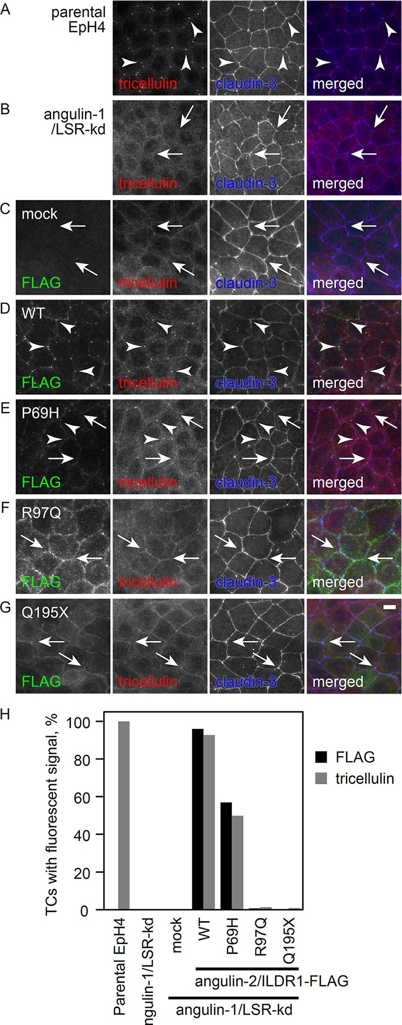
(A-G) Parental EpH4 cells, (A) LSR knockdown cells. (B) LSR knockdown cells stably expressing the mock FLAG vector (C) or wild-type (WT) human ILDR1. Immunostaining of (D) p.P69H, (E) p.R97Q, (F) or p.Q195X (G) was performed using anti-FLAG mAb (green), anti-tricellulin mAb (red) and anti-claudin-3 pAb (blue). Arrowheads and arrows indicate tricellular contacts with and without tricellulin localization, respectively. Scale bar: 10 μm. (H) Statistical analysis of FLAG and tricellulin localization at TCs. More than 600 TCs for each cell clone were examined for FLAG and tricellulin accumulation.