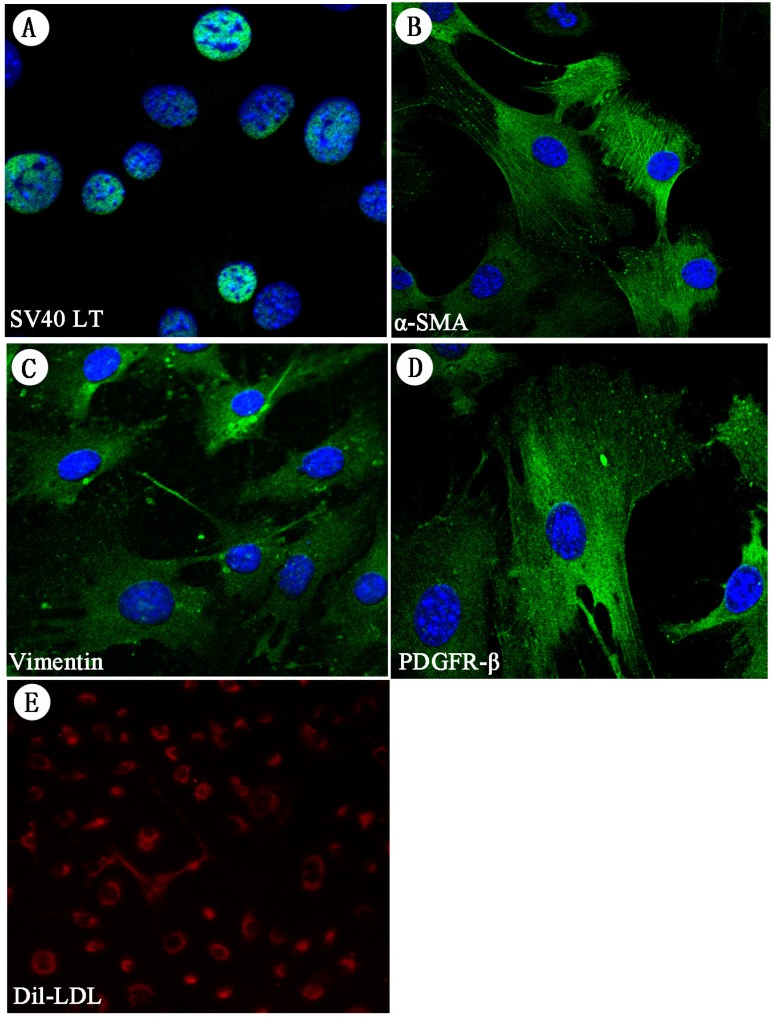
Figure 4.
Immunofluorescence staining of HSC-Li cells and DiI-LDL uptake. A. The expression of SV40LT proteins was positive in the HSC-Li cells. It was localized in the nucleus of HSC-Li cells. Cells were costained with DAPI to identify nuclei. Original magnification: ×400. B-D. The expression of α-SMA, vimentin, and PDGFR-β proteins was also positive in the HSC-Li cells. Cells were costained with DAPI to identify nuclei. Original magnification: ×400. E.The HSC-Li cells strongly endocytosed LDL after 24 h of culture with DiI-LDL. Original magnification: ×200.

