
Figure 4. Histochemistry images after sialidase treatment.
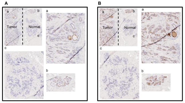
A. Staining with anti T-antigen antibody for breast tumor section with adjacent normal section. The section is labeled as a. Tumor section b. Ductal region in normal section c. Lobular section in normal section. B. Staining with PNA lectin for breast tumor section with adjacent normal section. The section is labeled identical to panel A.