Figure 6.
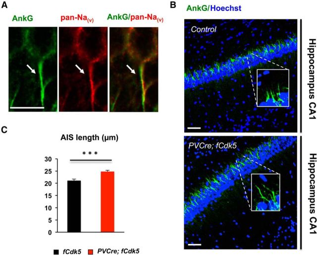
An increase in the length of the AIS in pyramidal neurons in the hippocampus of PVCre;fCdk5 mice. A, AnkG (green) colocalizes with pan-Nav (red) immunoreactivity at the AIS. B, AnkG immunoreactivity (green) as a marker of AIS of the pyramidal neurons in the corresponding hippocampal sections from control (fCdk5) and PVCre;fCdk5 animals (blue, Hoechst). Scale bars: A, 10 μm; B, 50 μm. C, Quantification of the length (micrometers) of the AIS in the neurons in the CA1 area of the hippocampus revealed by AnkG immunoreactivity. Magnification: main images, 20×; insets, 100×. Approximately 100 neurons were scored in the corresponding hippocampal sections from three pairs of control and PVCre;fCdk5 3.5-month-old littermate mice. Note a significant increase in the average length of the AIS in PVCre;fCdk5 mice compared with controls. Black, Control mice; red, PVCre;fCdk5 mice. ***p < 0.001; error bars indicate ±SEM.

