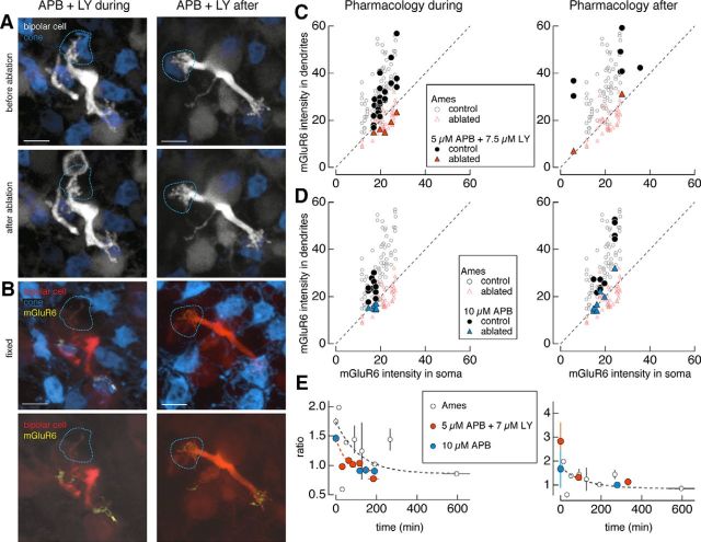
Figure 5.
Pharmacological occupation of glutamate receptors is insufficient to rescue mGluR6. A, B, Two-photon images of type 6 ON cone bipolar cells in live retina (A) and confocal images of the same bipolar cells in fixed retina (B). A, Type 6 ON cone bipolar cells in which the mGluR6 agonist l-APB (5 μm) and antagonist LY-341495 (7.5 μm) were perfused onto the retina (left) during and after or (right) after ablation of a cone (dotted lines). C, D, Average dendritic and somatic mGluR6 immunostaining intensities for control (circles) and ablated (triangles) cones under Ames solution (open symbols) and under 5 μm l-APB and 7.5 μm LY-341495 (C; closed symbols) or 10 μm l-APB (D; closed symbols) applied (left) during and after or (right) after the ablation of cones. Results from the one-way ANOVA for control (ablated) cones across the Ames solution and antagonist and/or agonist manipulations for the ratio of dendritic to somatic mGluR6 intensities in C [F(2,96) = 11.38, p = 3.7e-5 (F(2,50) = 1.4, p = 0.26)] and D [F(2,84) = 1.84, p = 0.17 (F(2,57) = 0.48, p = 0.62)]. E, Average ratio of dendritic to somatic intensities of mGluR6 immunostaining across the population of type 6 ON cone bipolar cells as a function of the interval between the ablation and fixation for bipolar cells under Ames solution (open black circles) or (left) during and after or (right) after 5 μm l-APB and 7.5 μm LY-341495 (orange closed circles) or 10 μm l-APB (blue closed circles). Data points represent mean ± SEM. Data points under APB fit with a single exponential with the following coefficients: y0 = 0.89 ± 0.14, A = 0.57 ± 0.16, τ = 50.6 ± 100. The time constant for receptor elimination (τ) is 51 min. Scale bars, 5 μm.

