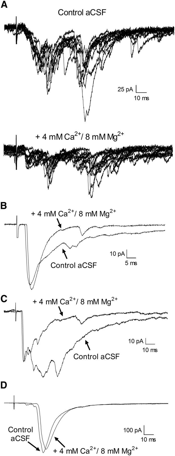
Figure 6.

Effect of high external divalent cations on primary afferent-evoked EPSCs in adult lamina I projection neurons. A, Top, Example of complex EPSCs that were evoked by primary afferent stimulation in the presence of normal aCSF. The EPSCs were classified as polysynaptic based on their variable onset latency and the presence of failures during repetitive stimulation (data not shown). Bottom, Bath application of an external solution containing 4 mm Ca2+ and 8 mm Mg2+ to suppress polysynaptic transmission significantly decreased the amplitude of these primary afferent-evoked EPSCs. B, C, Representative examples of EPSCs evoked in adult lamina I projection neurons that consisted of a short-latency component which was recruited at low-threshold A-fiber stimulus intensities (B, 20 μA; C, 30 μA) and followed high-frequency stimulation, in addition to longer-latency components. Perfusion with a high Ca2+/Mg2+ solution reduced the late phase of the EPSC without affecting the amplitude of the short-latency component, suggesting that this short-latency EPSC reflects a monosynaptic connection onto the projection neuron. A small delay in the EPSC latency (and the onset of the CAP in the dorsal root recordings; data not shown) was often observed, which may relate to the elevated firing threshold in the presence of high external divalents. D, Example of an EPSC evoked by the stimulation of low-threshold C-fibers (threshold = 20 μA) that was classified as monosynaptic based on its ability to follow repetitive stimulation (data not shown). Bath application of high Ca2+/Mg2+ had a minimal effect on the amplitude of the evoked EPSC, thus providing additional support of a direct connection between low-threshold C-fibers and mature projection neurons.
