Figure 7.

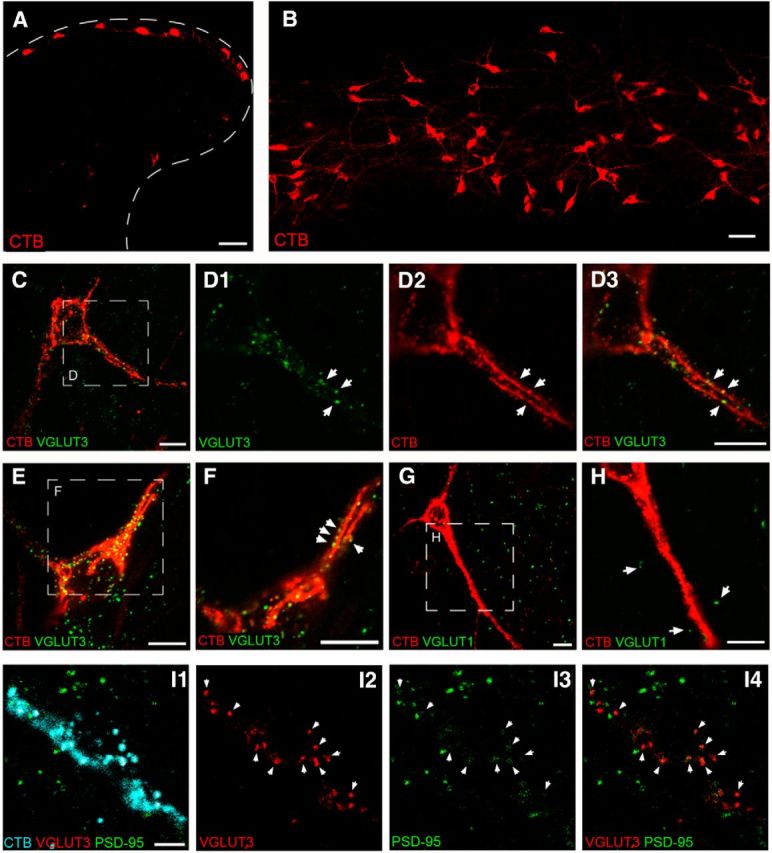
Adult lamina I projection neurons are innervated by VGLUT3-expressing axonal boutons. A, Transverse section of spinal cord illustrating lamina I neurons back-labeled via injections of CTB into the parabrachial nucleus. B, Horizontal (60 μm) section through the superficial dorsal horn demonstrating robust somatic and dendritic labeling of numerous ascending projection neurons. Scale bars: A, B, 50 μm. C, D, Immunohistochemical localization of the vesicular glutamate transporter VGLUT3, a known marker for low-threshold C-fiber mechanoreceptors, revealed VGLUT3-positive boutons (green) in close apposition (arrows) to the soma and dendrites of projection neurons identified by staining for CTB (red). C, Representative of a flattened projection of eight z-stack optical slices, whereas D1–D3 represents a single 0.5-μm-thick optical section shown at a higher-magnification from the boxed region in C. E, Flattened projection of nine z-stack optical slices illustrating another representative projection neuron which received numerous putative synaptic contacts from VGLUT3-containing afferents. F, Single 0.5 μm optical slice showing higher-magnification of the boxed region in E. G, In contrast, lamina I projection neurons (red) were rarely contacted by axonal boutons expressing VGLUT1 (green), a marker of large myelinated fibers associated with innocuous mechanoreceptors. H, Higher-magnification of the boxed region in G, demonstrating a lack of VGLUT1-positive boutons (arrows) in the immediate vicinity of the adult projection neuron. Scale bars, C–H, 10 μm. I, Single 0.5 μm optical sections illustrating the soma and proximal dendrites of a fusiform projection neuron back-labeled with CTB (cyan; I1) which receives input (arrows) from VGLUT3-IR boutons (red; I2, I4) at postsynaptic sites expressing PSD-95 (green, I3, I4). Scale bar, 5 μm.
