
Figure 8.
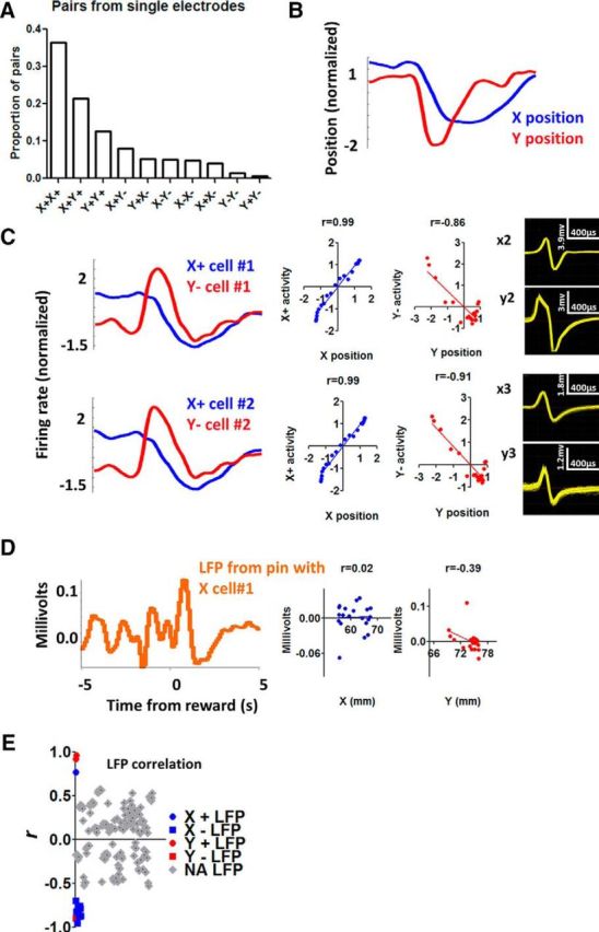
Single-unit activity from a single electrode compared with LFP signal from the same electrode. A, Probability of co-occurring neurons recorded from the same electrode. B, Illustration of the per-reward change in x- and y- coordinates during a single session. C, From the same session, 2 X+ neurons and 2 Y− neurons are shown with their waveforms. All cells show high correlation with position coordinates. Correlation analysis is performed on data from a 10 s perireward time window (5 s before and 5 s after reward delivery). D, LFP recorded from the same electrode as X+ cell #1. Correlation between LFP and position coordinates is much weaker. E, The distribution of r values for all the recorded LFP channels. Of the 166 LFP channels analyzed, only 15 showed significant correlation with position coordinates (colored, p < 0.001). Thus, when activity is summed from different types of neurons, the resulting correlation between neural activity and position is weaker. The relatively rare examples of LFP being highly correlated with position coordinates suggest that neurons belonging to a particular class (e.g., X+) might be located close to each other.