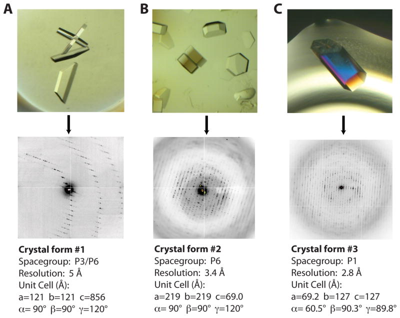
Figure 1. Three Rho crystal-forms obtained from substrate matrix screening.
A) Crystal-form I (top) grew as thin hexagonal plates that diffract to relatively low resolution. A close-up view of the diffraction pattern (bottom) reveals the long unit cell edge that caused severe overlaps and prevented collection of a complete dataset.
B) Crystal-form II grew as thick hexagonal “lug-nuts” (top). Diffraction patterns revealed a P6 packing arrangement. Usable diffraction maxima extended to 3.4 Å resolution.
C) Crystal-form III grew as large hexagonal rods free from any obvious morphological defects (top). While the diffraction pattern is highly mosaic, these crystals diffracted to the highest resolution of any of the forms.

