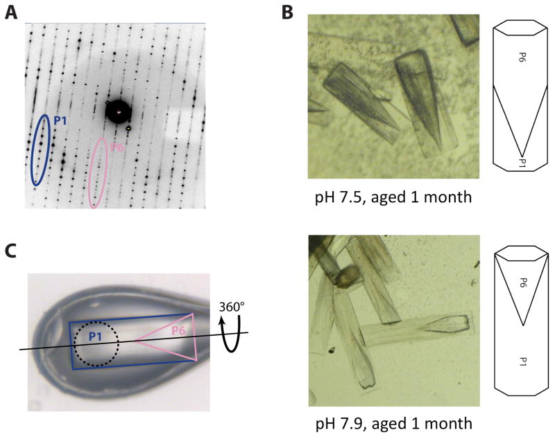
Figure 3. Overcoming a vexing form of coincident non-merohedral twinning.
A) Initial diffraction patterns obtained from crystal-form III (Figure 1C), revealed two distinct lattices. While the P1 diffraction dominated the pattern (blue/dark grey), a P6 diffraction pattern corresponding to crystal-form II was present at low resolution (pink/light grey). Since crystal-forms I and II shared a coincident ~69 Å unit cell edge, the diffraction patterns almost perfectly overlapped and prevented accurate measurement of low resolution P1 data.
B) Aging caused the P1 portion of each crystal to degrade at a faster rate than the P6 portion. When viewed under bright-field illumination, the two twin domains are clearly distinguishable. The P6 twin domain forms at the nucleating end of the crystals and has a “cone-shaped” protrusion that interleaved with the P1 twin domain (top). An X-ray beam aimed at the P1 tip of the crystal thus passes through a small fraction of the P6 twin domain, producing P6 diffraction only at low resolution as seen in panel A. Increasing the pH of crystal growth increased the relative size of the P1 twin domain (bottom).
C) Crystals grown at a pH of 7.9 were mounted in bendable cryo-loop and oriented such that the axis of crystal rotation (dashed line) was parallel with the long axis of the hexagonal rods. A 100 μM collimated beam (dashed circle) was directed into the P1 tip (blue/dark grey) of the crystal. Data collection in this orientation avoided contributions from the P6 twin domain (pink/light grey).

