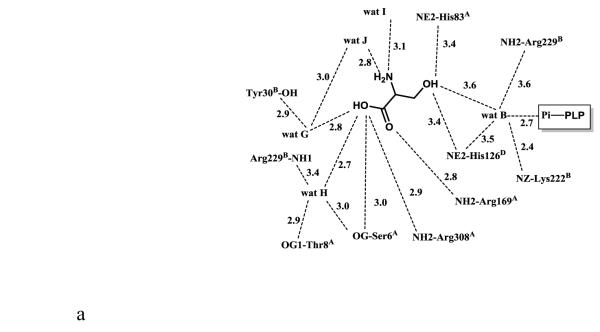
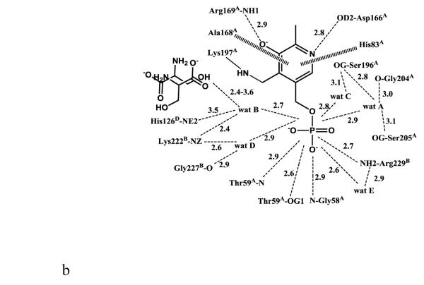
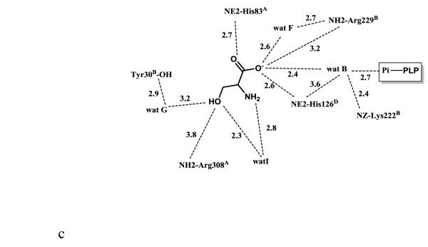
Figure 2.
Scheme of two-dimensional contacts between ligands, protein residues and the structural water molecules discussed in the text. Dotted lines indicate hydrogen-bond interactions and their length (in Å), and broad dashed lines represent hydrophobic interactions. (A) PLP contacts. (B) L-serine (with the carboxylate facing the arginines) contacts. (C) L-serine (with the carboxylate facing the histidines) contacts.


