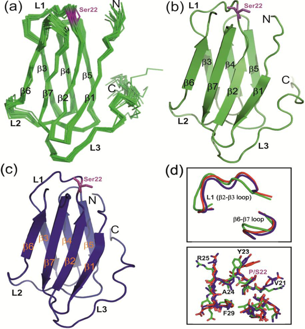
Fig. 7.
NMR and crystal structures of hCS(P22S). (a) Ensemble of 20 lowest energy solution NMR structures of hCS(P22S). (b) The lowest energy structure of hCS(P22S) in cartoon representation. (c) Crystal structure of hCS(P22S). All the secondary structure elements are marked. Ser22 is shown in magenta in stick representation. (d) Comparison of L1 loops in hCS (crystal, red), hCS(P22S) (NMR, green), and hCS(P22S) (crystal, blue) in backbone tube (top) and stick (bottom) representation. Residues in L1 loop are marked.

