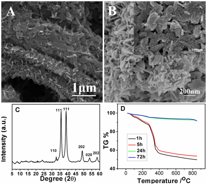
Figure 3.
Low-magnification SEM image (A) and high-magnification SEM image (B) of CuO superstructure prepared by immersing six-prismatic Cu-MOFs in 0.1 M NaOH for 72 h. Inset in B: partial enlargement. (C) XRD of the six-prismatic Cu-MOFs immersed in 0.1 M NaOH for 72 h. (D) TGA curves of complexes synthesized by immersing six-prismatic Cu-MOFs in 0.1 M NaOH for 1 h, 5 h, 12 h and 24 h.

