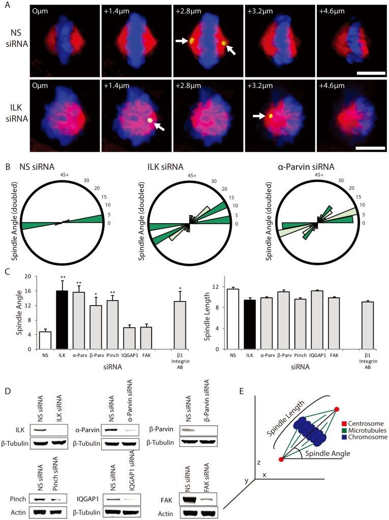
Figure 1. ILK siRNA causes the mitotic spindle to be misoriented.
A) Z-sections of non-specific and ILK siRNA transfected metaphase HeLa (Kyoto) cells showing centrosome location (Pericentrin; green/yellow), spindle shape (Tubulin; red) and DNA (Hoechst; blue). Arrows point to centrosomes. Distance from bottom of the cell is denoted on each figure in microns. B) Spindle angle distribution graphs of siRNA treated HeLa cells. C) Measurements of spindle angle along the Z-axis and spindle length in siRNA treated or β1-Integrin inhibitory antibody treated cells. n:30 for each condition. NS denotes non-specific siRNA control. D) Western blots of lysates from siRNA transfected cells demonstrating knock-down of the proteins studied. Western blots were cropped as indicated (black boxes). For all samples, SDS-PAGE and Western blotting were performed under the same experimental conditions. E) Diagram of mitotic spindle angle and length measurements. Scale bar 10 μm. * = p < 0.05; ** = p < 0.01. Error bars represent SEM.

