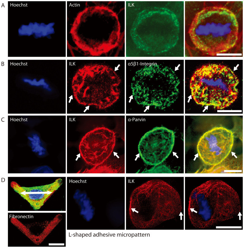
Figure 2. Localization of ILK, Actin, α5β1-Integrin and α-Parvin at the basal lamina of metaphase HeLa (Kyoto) cells.
A) ILK localizes to an Actin ring at the bottom of metaphase cells. B) ILK colocalizes with active α5β1-Integrin at the cell edge of metaphase cells (arrows). C) α-Parvin colocalizes with ILK at a ring on the bottom of a metaphase cell (arrows). D) First panels: Interphase HeLa cell adhering to an L-shaped micropattern. ILK (green), Hoechst (blue) and Fibronectin (red). For orientation, the double-headed arrow denotes the direction of tension when the cell enters mitosis. Other panels: ILK and Hoechst staining of metaphase cells grown on L-shaped micropatterned coverslips. ILK aligns with the long axis of the adherent cell, in the direction of tension that aligns the mitotic spindle (arrows). Scale bar 10 μm.

