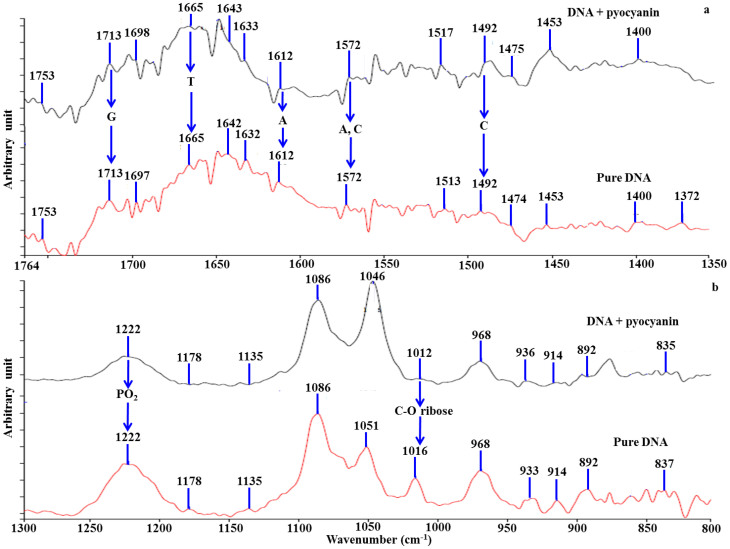
Figure 2. FTIR study on DNA-pyocyanin binding.
ATR-IR spectra of pure DNA and a pyocyanin-DNA mixture are shown. The addition of pyocyanin to DNA solutions led to a major change in intensity or shift of the peaks at 1665, 1572 and 1492 cm−1 for thymine (T), adenine (A) and cytosine (C) respectively (a). Pyocyanin interaction with DNA phosphate (PO2−) and ribose backbone are demonstrated by the change in intensity or shift of the peaks at 1178, 1135, 1016, 914 and 837 cm−1. The B-conformation of DNA was not changed by addition of pyocyanin as indicated by the absence of significant changes at peaks 1222 and 1086 cm−1 (b).

