
Figure 3. Neuroprotection of the compounds against the Aβ42-induced toxicity in cortical neurons (CN).
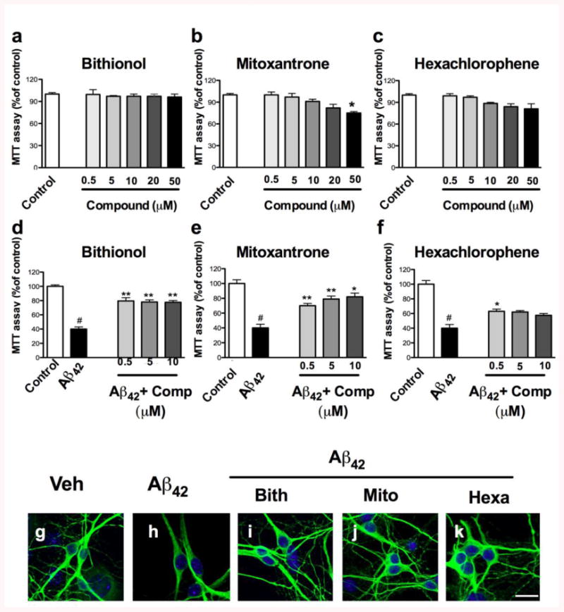
a-b) Cortical neurons were treated with different concentrations of the compounds (0.5, 5, 10, 20 and 50 μM) for 24 hours. d-f) The neurons were treated with 40 μM of the crude preparation of Aβ42 or co-treated with the crude preparation and the compounds at different concentrations (0.5, 5 and 10 μM) at 7 DIV. The effects of the compounds and the neuroprotection against the Aβ42-induced toxicity were evaluated using an MTT assay, and the control treatment was set to 100%. The error bars represent the mean±S.E. We used # to indicate the data compared to the control and * compared to the treatments with crude Aβ42 preparation, *p<0.05, **p<0.01, g-k). The neuroprotection of the compounds against Aβ42-induced toxicity was evaluated by staining the neurons with MAP2 (green) by immunofluorescence, and the nuclei were stained with DAPI (blue).