
Figure 6. Immunoblot analysis of Aβ species, ELISA test and γ-secretase activity in APPtg mice treated with bithionol, mitoxantrone and hexachlorophene.
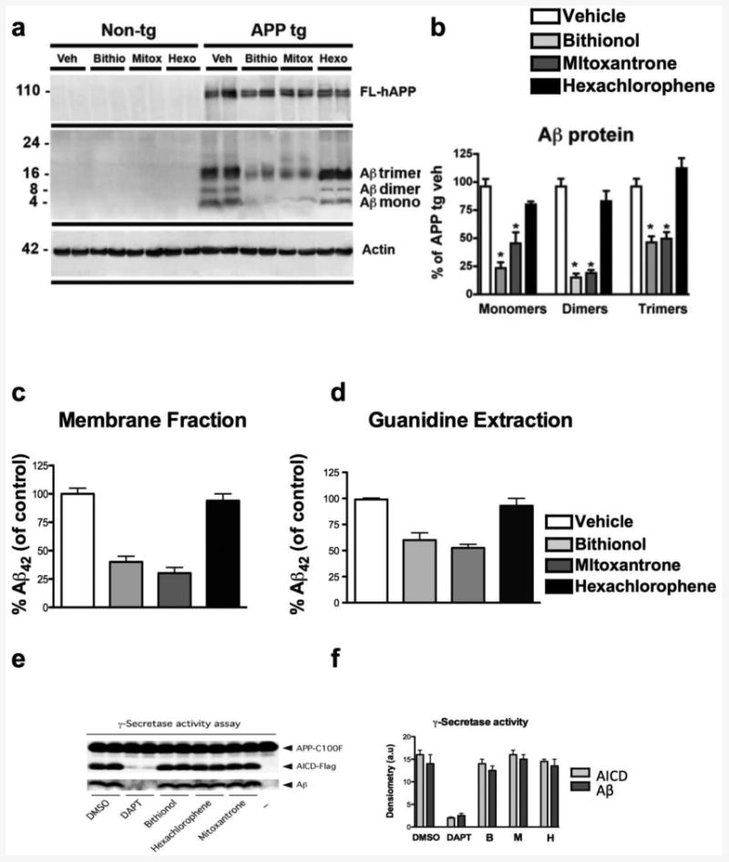
a)The mouse posterior brain tissues were homogenized in Buffer A, and the membrane fraction was probed with antibodies against Aβ (82E1) and β-actin. b) Semi-quantitative densitometric analyses of the bands representing Aβ monomers (4 kDa), dimers (8 kDa), and trimers (12 kDa). N=5 cases per group, *p<0.05 compared with APP/tg mice by ANOVA with post-hoc Dunnett's test. c) and d) quantitative analysis of Aβ42 levels in APPtg mice treated with bithionol, mitoxantrone and hexachlorophene by ELISA. The quantification of Aβ42 levels in the membrane fraction (c) and guanidine-HCl extracted samples (d) N=5 cases per group, female gender. *p<0.05 compared with APPtg mice by ANOVA with post-hoc Dunnett's test.
e-f) the compounds do not affect the processing of APP-C100-Flag and Aβ production by purified γ-secretase. γ-Secretase solubilized in 0.2% CHAPSO-HEPES, pH 7.5, was incubated at 37°C for 4 h with 1 μM C100-Flag substrate, 0.1% PC, 0.025% PE and 10 μM of bithionol, hexachlorophene and mitoxantrone. Activity assays included control reactions with 10 μM of the γ-secretase inhibitor DAPT, DMSO (1.6% w/v) or a reaction with omitted γ-secretase (-). The reactions were halted by adding 0.5% SDS, and the resulting products were detected with anti-Aβ and -AICD-Flag antibodies 6E10 and M2, respectively. The relative levels of AICD-Flag or Aβ were estimated by densitometry (mean±SD; n=2).