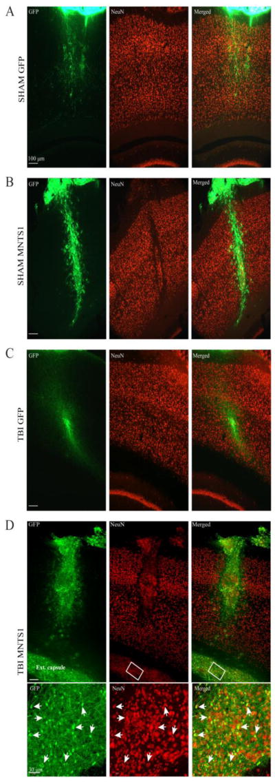
Figure 1.
GFP-positive cell survival and neuronal differentiation 6 weeks post surgery. A-D) Representative images showing GFP-positive transplanted NPCs from a sham/GFP, sham/MNTS1, TBI/GFP, and a TBI/MNTS1 animal, respectively. B, D) Several MNTS1-transduced NPCs were also immunoreactive for NeuN, indicating neuronal differentiation. D) After TBI, numerous MNTS1-transduced NPCs were observed ventrolateral from needle tract and within the external capsule. Boxed region in D demarcates area of high magnification in bottom panel. Arrows indicate transplanted NPCs with GFP/NeuN double-labeled immunoreactivity. Green, GFP; red, NeuN; ext. capsule, external capsule.

