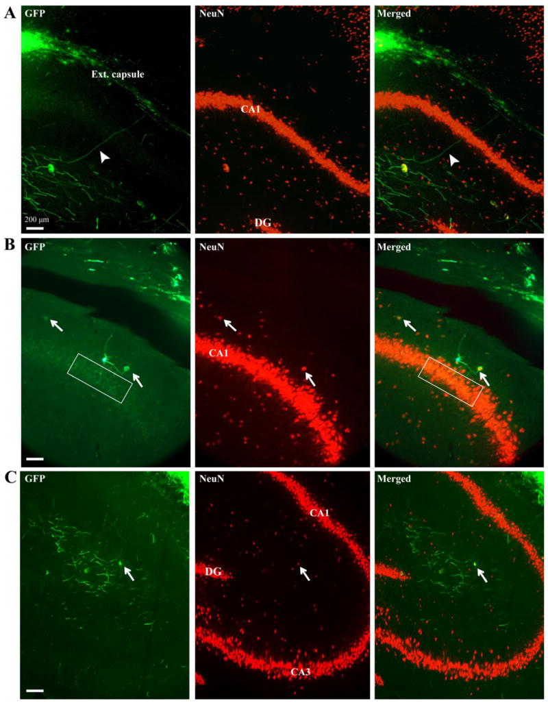
Figure 5.
GFP-positive processes and some GFP/NeuN-positive cell bodies infiltrated the hippocampus in both injured and non-injured animals 6 weeks post surgery. A) NPC grafts residing in external capsule extended GFP-positive processes (arrowhead) through CA1 of the hippocampus and into the stratum radiatum. B) GFP-positive cell bodies, one of which was also NeuN-positive (arrow), extended processes into CA1 of the hippocampus (boxed region). C) GFP-positive processes and GFP/NeuN-positive cell body (arrow) were observed within the stratum radiatum. Green, GFP; red, NeuN; ext. capsule, external capsule; DG, dentate gyrus; CA, cornu Ammonis.

