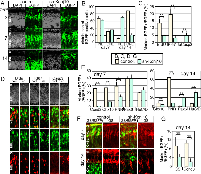
Figure 1.
Expression of sh-Kcnj10 in mouse retinal explants. A: Control-enhanced green fluorescent protein (EGFP) or sh-Kcnj10/EGFP expression plasmids were electroporated into isolated retinas at E18, and cultured as explants on the indicated days. B: Sub-retinal distribution of EGFP-positive cells was calculated on days 7 and 14. C, D: Proliferated and apoptotic cells were analyzed after 3 days of culture with immunostaining. The population of the immunostaining signal-positive cells (C) and immunostained pattern (D) are shown. E–H: Differentiation was examined with immunostaining of markers for retinal subpopulation. Calculation of the population of marker and EGFP double-positive cells in the total EGFP positive cells (E, G). The number of marker-positive cells in each layer was counted in 150-μm-wide retina (F). Staining patterns of GS and EGFP (H) are shown. Nuclei were visualized with 4',6-diamidino-2-phenylindole (DAPI) staining (gray) in A, D. P value; * <0.05, **<0.01 (the Student t test from at least three independent samples). cont, control; sh, sh-Kcnj10; ONL, outer nuclear layer; INL, inner nuclear layer; ONBL, outer neuroblastic layer; GCL, ganglion cell layer; aCasp3, active caspase 3; GS, glutamine synthetase.

