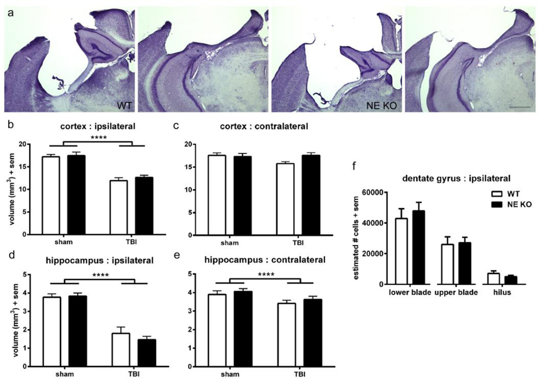
Figure 3. NE gene deficiency does not preserve injury-induced tissue damage long-term.
Stereological analysis was performed on cresyl violet stained sections to assess atrophy in the dorsal cortex and hippocampus at 3 months of age (~ 2 months post-injury). Representative images are presented from WT and NE KO brains both anteriorly and posteriorly (a; scale bar =500 µm). Quantification revealed a reduction of cortical volume in TBI mice compared to sham controls (2-way ANOVA effect of injury, ****p<0.0001), which was similar in WT and NE KO mice (b). Contralateral cortex volume was not affected by either injury or genotype (c). Hippocampal volumes were affected by injury both ipsilateral and contralateral to the impact site (d; 2-way ANOVA effect of injury ****p<0.0001), to a similar extent in both WT and NE KO mice. The surviving neuronal population in specific regions of the ipsilateral hippocampus DG (e) was calculated using the optical fractionator method. Consistent with volumetric analyses, no differences were observed between WT and NE KO mice (t-tests n.s). n=10–12/group.

