
Figure 1.
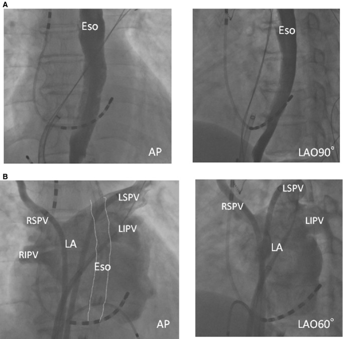
Representative esophagraphy and pulmonary venography. A, Esophagrams reveal the esophageal courses. B, Pulmonary venography shows the anatomy of the PV antrum. AP indicates anteroposterior view; Eso, esophagus; LA, left atrium; LAO, left oblique view; LIPV, left inferior PV; LSPV, left superior PV; PV, pulmonary vein; RIPV, right inferior PV; RSPV, right superior PV.