Figure 1.

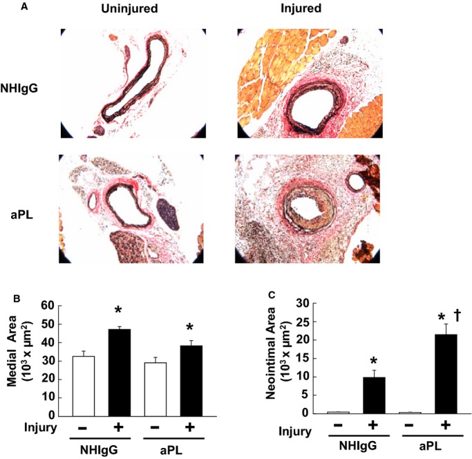
Antiphospholipid antibodies (aPLs) promote carotid artery neointima formation after endothelial denudation in mice. A, Male wild‐type mice were treated with human IgG from healthy nonautoimmune individuals (NHIgG; 100 μg/mouse) or aPLs (100 μg/mouse) 1 day before carotid artery endothelial denudation by using an epon‐resin probe and every other day after denudation for 2 weeks. Medial hypertrophy and neointima formation were then evaluated by histologic analysis. Representative photomicrographs are shown of uninjured (left panels) and injured carotid arteries (right panels). B, Medial area was calculated as the area encircled by the external elastic lamina minus the area encircled by the internal elastic lamina. Values are mean±SEM. For the NHIgG‐treated group (n=10), median was 30 396 (uninjured) and 45 186 (injured). For the aPL‐treated group (n=9), median was 26 861 (uninjured) and 37 477 (injured). C, Neoinimal area was calculated by subtracting the luminal area from the area encircled by the internal elastic lamina. Values are mean±SEM. For the NHIgG‐treated group (n=10), median was 387 (uninjured) and 7974 (injured). For the aPL‐treated group (n=9), median was 288 (uninjured) and 18 226 (injured). *P<0.05 vs uninjured, †P<0.05 vs NHIgG.
