
Figure 7.
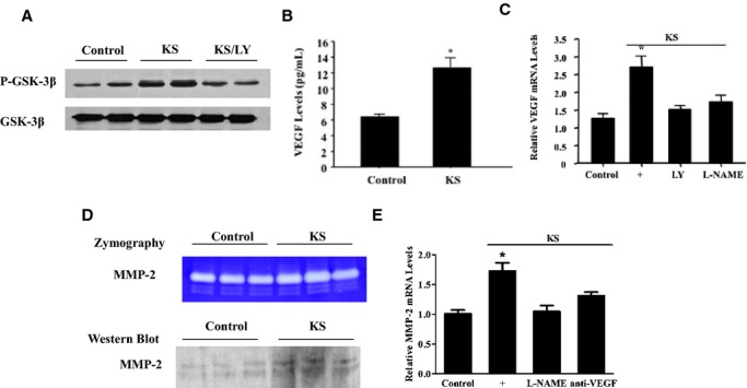
KS increases GSK‐3β phosphorylation as well as VEGF and MMP‐2 levels via phosphoinositide 3‐kinase–Akt pathways in EPCs. A, Western blot of phosphorylated and total GSK‐3β. B and C, Protein (B) and mRNA (C) levels of VEGF. D and E, Zymography and Western blot (D) and mRNA (E) levels of MMP‐2. *P<0.01 vs other groups. Data are expressed as mean±SEM, n=3. EPCs indicates endothelial progenitor cells; GSK‐3β, glycogen synthase kinase‐3β; KS, kallistatin; LY, LY294002; MMP‐2, matrix metalloproteinase‐2; VEGF, vascular endothelial growth factor.