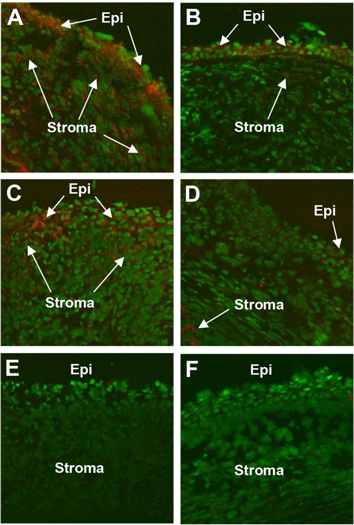
Figure 2. HMGB1 corneal staining after VIP treatment.
More positive staining for HMGB1 (red) was seen after PBS (A) when compared to VIP treatment (B) at 1 day p.i. Staining at 5 days p.i. also showed more corneal HMGB1 (red) in the PBS (C) vs VIP treated animals (D). Negative controls in which species specific IgG replaced the primary Ab are negative for HMGB1 staining (red) after PBS (E) and VIP (F) treatments. (Epithelium=Epi and stroma=Stroma). Mag=115X (n=5/group/time/assay)

