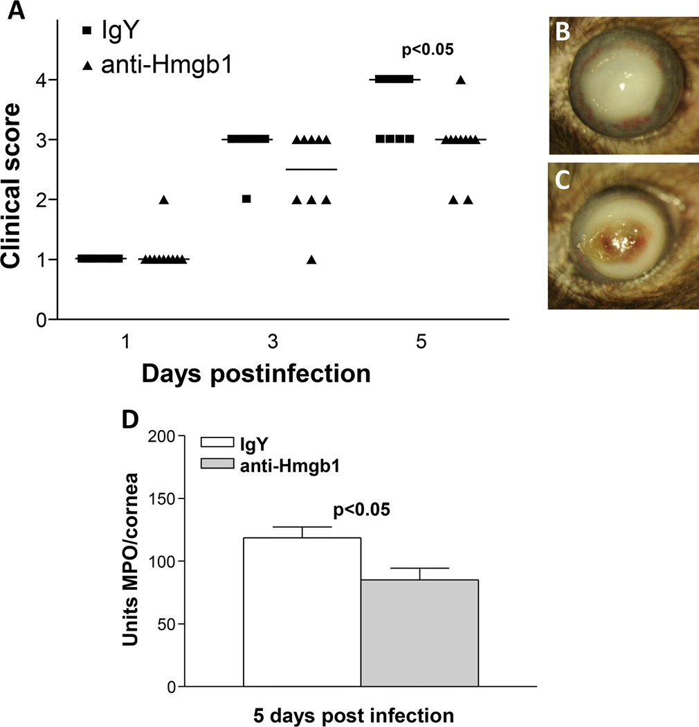
Figure 8. HMGB1 neutralization.
Clinical score (A) shows that antibody neutralization of HMGB1 resulted in significantly less disease at 5 days p.i. (p<0.05) with no difference in disease scores seen at 1 and 3 days p.i. (n=10/group/time). Photographs taken with a slit lamp of representative corneas at 5 days p.i. show opacity with no perforation in the cornea of anti-HMGB1 treated mice (+3) (B) and corneal perforation in the IgY treated control cornea (+4) (C). MPO assay (D) detected fewer PMN in the cornea of anti-HMGB1 vs IgY control treated mice at 5 days p.i. (p<0.05) (n=10/group/time for each assay) Clinical score data (A) analyzed using a non-parametric Mann-Whitney test, with medians indicated for each group. Data shown in (D) analyzed using a two-tailed Student’s t-test and shown as the mean ± SEM. B and C Mag= 5X.

